Cervical Cells
Home / Microscopic / Histopathology / Cervical Cells
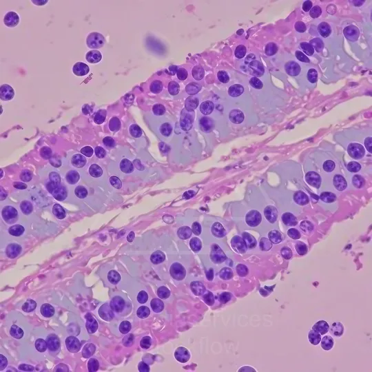
This collection contains 11 assets available in multiple resolutions and aspect ratios.
1 Micro 12 Pathology 14 Histopathology Cervical Cells
Resolution: 11095x6048 | Format: 16:9

Cervical epithelial cells with koilocytotic changes indicative of HPV infection.
⬇ Download / Buy on Stockflow.media
2 Micro 12 Pathology 14 Histopathology Cervical Cells
Resolution: 8192x8192 | Format: 1:1
Histopathology image of cervical epithelial cells.
⬇ Download / Buy on Stockflow.media
3 Micro 12 Pathology 14 Histopathology Cervical Cells
Resolution: 11095x6048 | Format: 16:9

Histopathology slide of cervical epithelial cells showing squamous cells with nuclear changes and koilocytosis.
⬇ Download / Buy on Stockflow.media
4 Micro 12 Pathology 14 Histopathology Cervical Cells
Resolution: 8192x8192 | Format: 1:1

Histopathology image of cervical squamous cells showing nuclear enlargement with atypia and koilocyte-like features.
⬇ Download / Buy on Stockflow.media
5 Micro 12 Pathology 14 Histopathology Cervical Cells
Resolution: 11095x6048 | Format: 16:9

Histopathology micrograph of cervical cells with numerous small round purple nuclei scattered in a pink-stained background.
⬇ Download / Buy on Stockflow.media
6 Micro 12 Pathology 14 Histopathology Cervical Cells
Resolution: 8192x8192 | Format: 1:1

Histopathology of cervical cells.
⬇ Download / Buy on Stockflow.media
7 Micro 12 Pathology 14 Histopathology Cervical Cells
Resolution: 11095x6048 | Format: 16:9

Histopathology of cervical cells showing koilocytes and epithelial cell changes.
⬇ Download / Buy on Stockflow.media
8 Micro 12 Pathology 14 Histopathology Cervical Cells
Resolution: 8192x8192 | Format: 1:1

Histopathology image of cervical squamous cells showing koilocytosis and HPV-associated epithelial changes.
⬇ Download / Buy on Stockflow.media
04 Histopathology
Resolution: 2160x2160 | Format: 1:1
Histopathology image of cervical cells at micro level, displaying numerous purple-stained nuclei embedded in fibrous tissue, illustrating cellular morphology and tissue architecture characteristic of a cervical lesion.
⬇ Download / Buy on Stockflow.media
5 Histopathology
Resolution: 3840x21 | Format: 16:9
Histopathology micrograph of cervical cells showing scattered nuclei and pink-stained connective tissue, highlighting cellular composition and structural details typical of cervical tissue at high magnification.
⬇ Download / Buy on Stockflow.media
6 Histopathology
Resolution: 2160x3840 | Format: 9:16
Histopathology slide of cervical cells showing layered epithelial debris and dark-staining nuclei amid pink connective tissue, highlighting cellular details and inflammatory features characteristic of cervical cytology.
⬇ Download / Buy on Stockflow.media