Cellular
Home / Microscopic / Cellular / Cellular
This collection contains 113 assets available in multiple resolutions and aspect ratios.
1 Micro 14 Cellular
Resolution: 11092x6050 | Format: 16:9
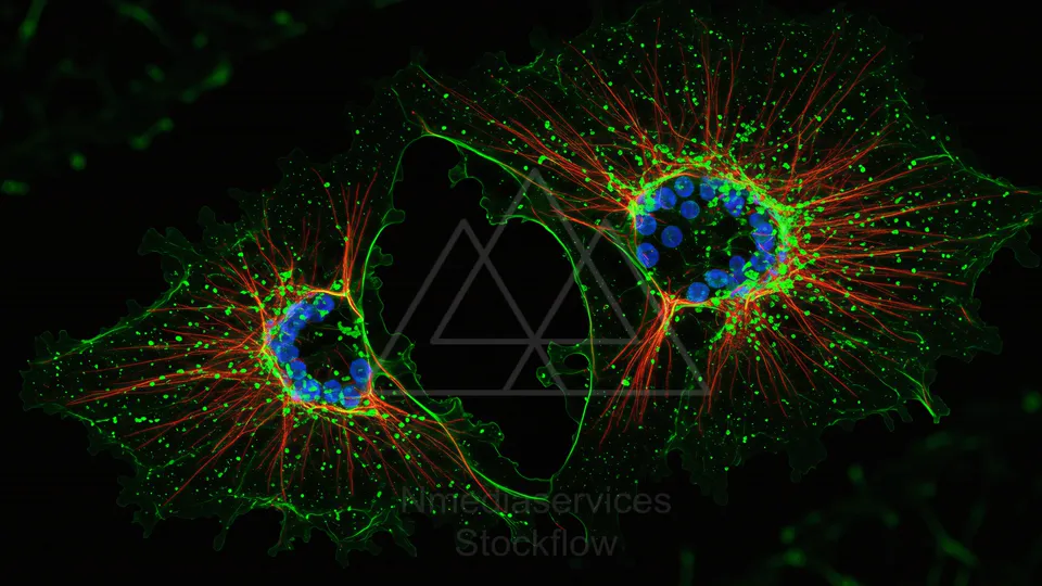
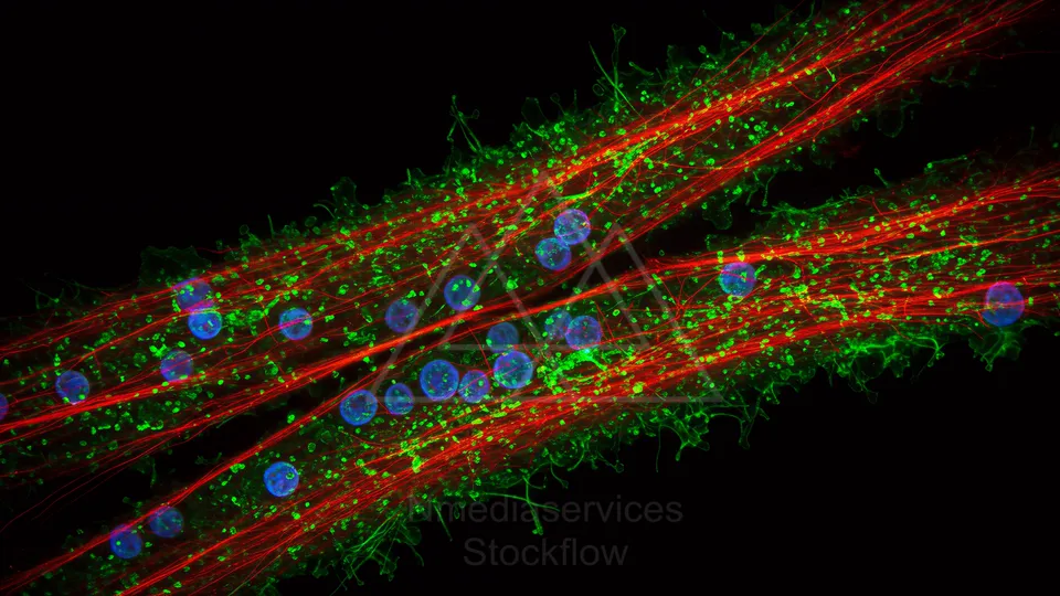
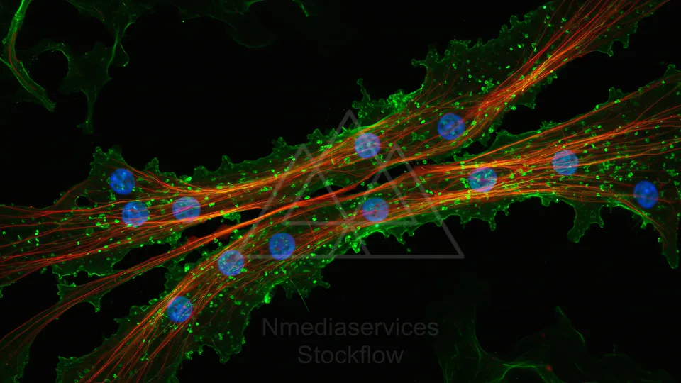
Fluorescent micrograph of two adjacent cells showing a green cytoskeleton and membrane, red filamentous networks, and blue nuclei.
⬇ Download / Buy on Stockflow.media
2 Micro 14 Cellular
Resolution: 8192x8192 | Format: 1:1
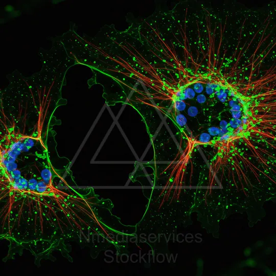
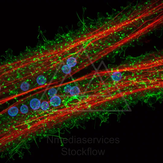
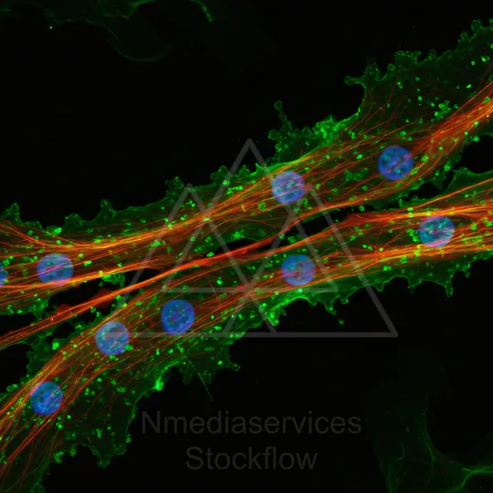
Fluorescence micrograph of a cultured cell showing green membrane/cytoskeleton, red actin fibers, and blue nuclei.
⬇ Download / Buy on Stockflow.media
3 Micro 14 Cellular
Resolution: 6050x11092 | Format: 9:16
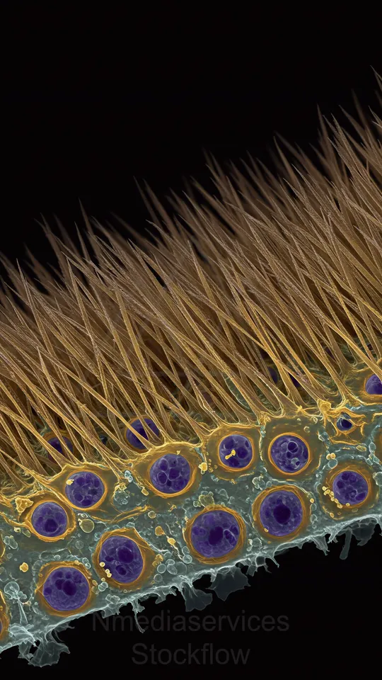
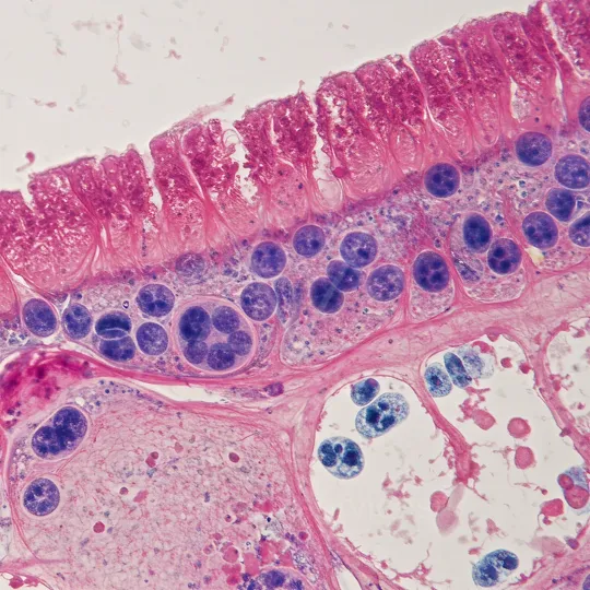
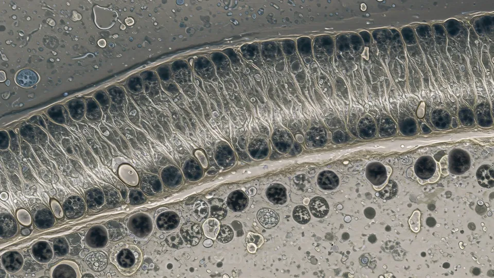
False-colored microscopic cross-section of ciliated epithelial cells, showing a row of hairlike cilia above rounded cells with prominent nuclei.
⬇ Download / Buy on Stockflow.media
4 Micro 14 Cellular
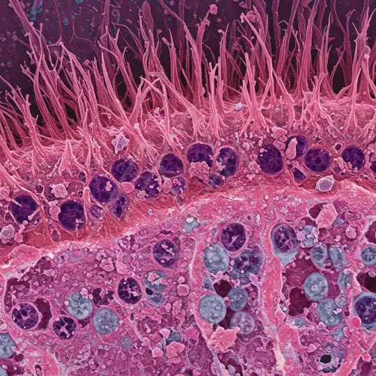
Resolution: 11092x6050 | Format: 16:9

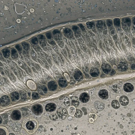
False-colored microscopic cross-section of ciliated epithelial cells, showing a row of hairlike cilia above rounded cells with prominent nuclei.
⬇ Download / Buy on Stockflow.media
5 Micro 14 Cellular
Resolution: 8192x8192 | Format: 1:1
False-colored microscopic cross-section of ciliated epithelial cells, showing a row of hairlike cilia above rounded cells with prominent nuclei.
⬇ Download / Buy on Stockflow.media
6 Micro 14 Cellular
Resolution: 11092x6050 | Format: 16:9
False-colored microscopic cross-section of ciliated epithelial cells, showing a row of hairlike cilia above rounded cells with prominent nuclei.
⬇ Download / Buy on Stockflow.media
7 Micro 14 Cellular
Resolution: 8192x8192 | Format: 1:1
False-colored microscopic cross-section of ciliated epithelial cells, showing a row of hairlike cilia above rounded cells with prominent nuclei.
⬇ Download / Buy on Stockflow.media
1 Micro 14 Cellular
Resolution: 11092x6050 | Format: 16:9
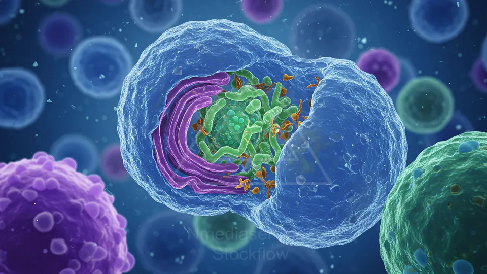
Colorful 3D render of a cell with its inner organelles, featuring green mitochondria and purple membranous structures.
⬇ Download / Buy on Stockflow.media
2 Micro 14 Cellular
Resolution: 8192x8192 | Format: 1:1
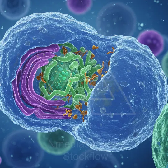
Colorful 3D render of a cell with its inner organelles, featuring green mitochondria and purple membranous structures.
⬇ Download / Buy on Stockflow.media
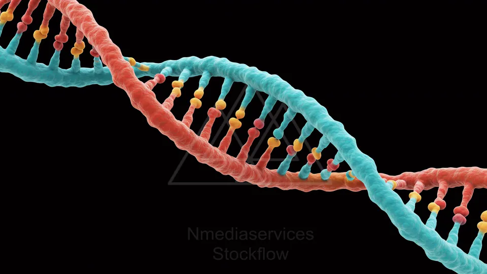
3 Micro 14 Cellular
Resolution: 11092x6050 | Format: 16:9
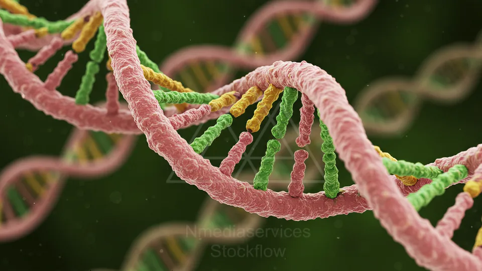
Close-up of a colorful DNA double helix with pink, green, and yellow strands against a dark background.
⬇ Download / Buy on Stockflow.media
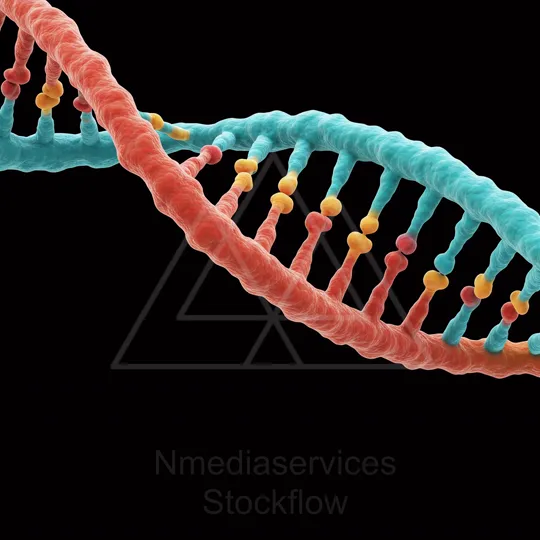
4 Micro 14 Cellular
Resolution: 8192x8192 | Format: 1:1
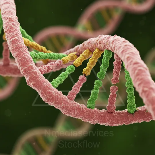
Close-up illustration of a DNA double helix with pink and green strands intertwined, highlighting the molecular structure.
⬇ Download / Buy on Stockflow.media
5 Micro 14 Cellular
Resolution: 11092x6050 | Format: 16:9
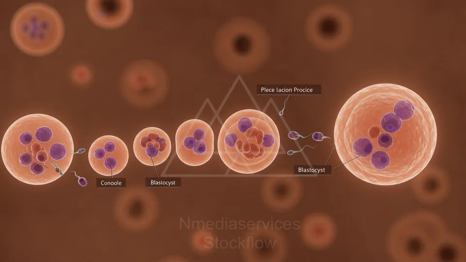
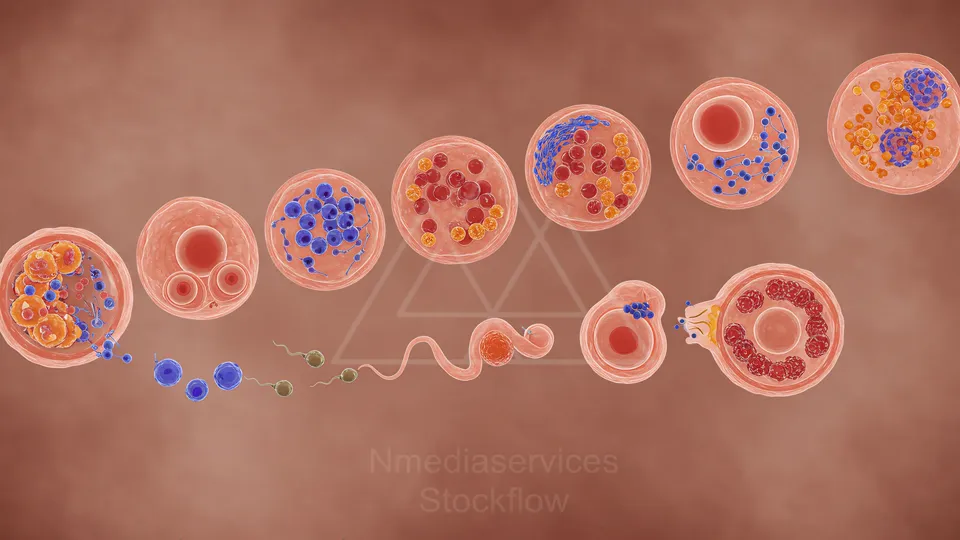
Illustration of early embryonic development from fertilization through cleavage to a blastocyst.
⬇ Download / Buy on Stockflow.media
1 Micro 14 Cellular
Resolution: 6050x11092 | Format: 9:16

Illustration of a fetus in the womb with a detailed cross-section of the placenta and surrounding tissues.
⬇ Download / Buy on Stockflow.media
2 Micro 14 Cellular
Resolution: 6050x11092 | Format: 9:16
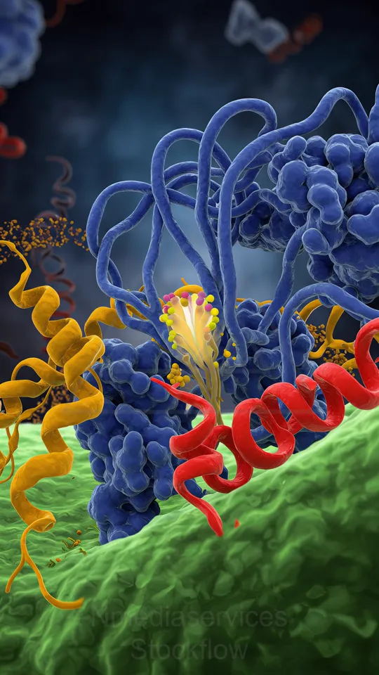
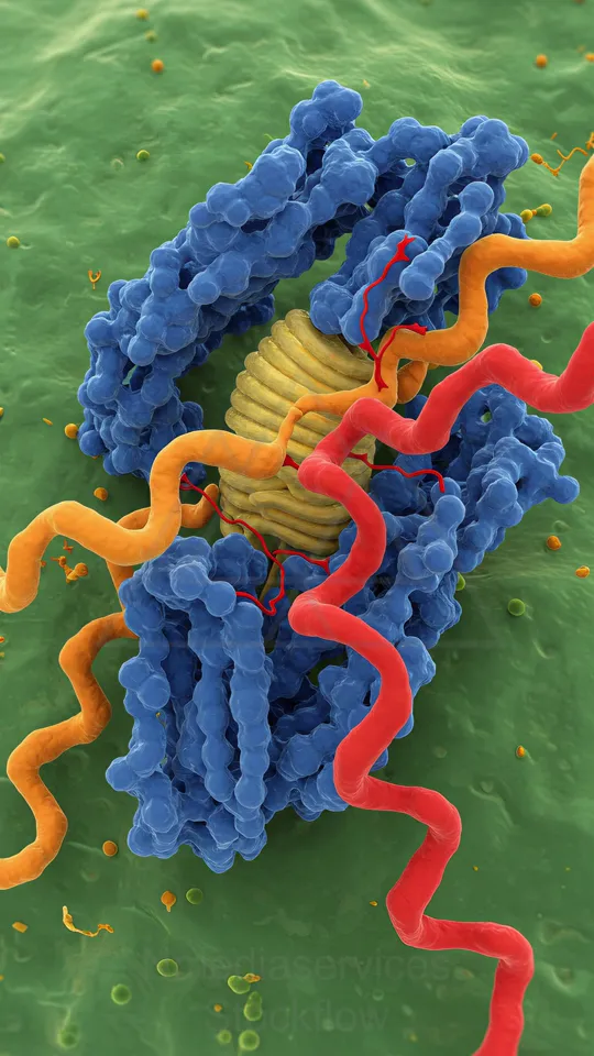
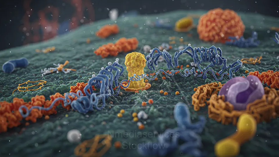
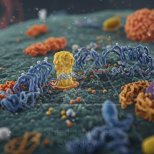
Colorful high-midelity illustration of a cell membrane with blue filament proteins, red helices, yellow signaling molecules, and a green lipid bilayer.
⬇ Download / Buy on Stockflow.media
3 Micro 14 Cellular
Resolution: 11092x6050 | Format: 16:9
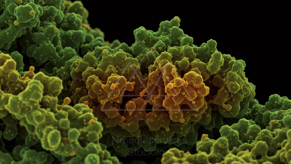
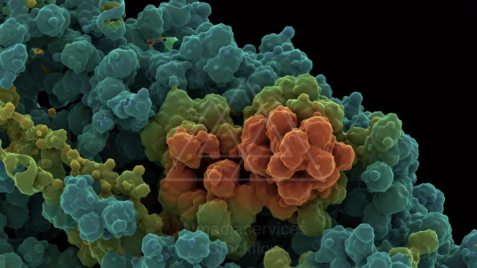
Colorized 3D rendering of clustered cellular structures in green and yellow hues.
⬇ Download / Buy on Stockflow.media
4 Micro 14 Cellular
Resolution: 8192x8192 | Format: 1:1
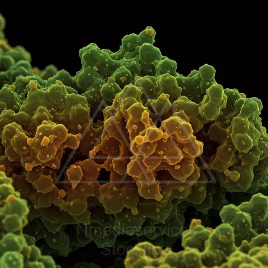
Colorized electron microscopy image of clustered cells.
⬇ Download / Buy on Stockflow.media
5 Micro 14 Cellular
Resolution: 11092x6050 | Format: 16:9
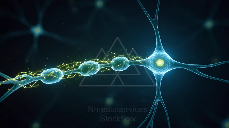
Blue neural network with glowing synapses connecting spherical nodes.
⬇ Download / Buy on Stockflow.media
6 Micro 14 Cellular
Resolution: 8192x8192 | Format: 1:1
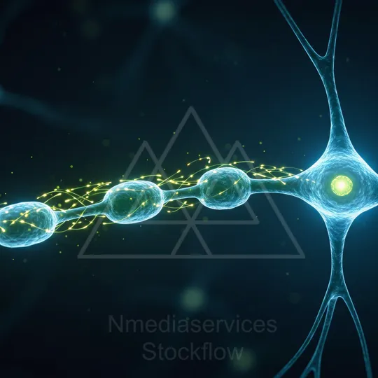
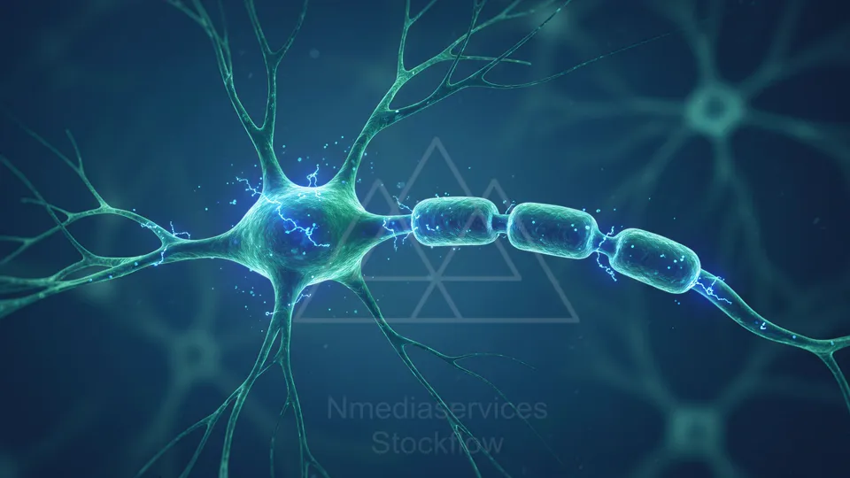
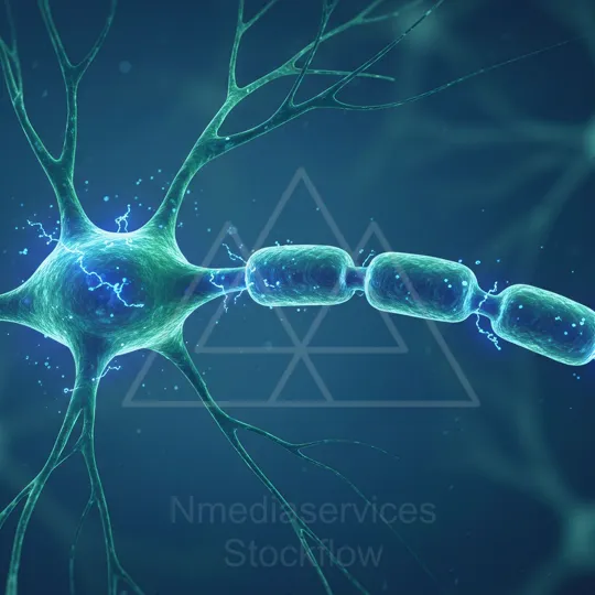
A detailed illustration of a neuron with an axon and glowing synapses, featuring neon blue cellular structures and bright yellow neural signals.
⬇ Download / Buy on Stockflow.media
7 Micro 14 Cellular
Resolution: 6050x11092 | Format: 9:16
A detailed illustration of a neuron with an axon and glowing synapses, featuring neon blue cellular structures and bright yellow neural signals.
⬇ Download / Buy on Stockflow.media
8 Micro 14 Cellular
Resolution: 6050x11092 | Format: 9:16
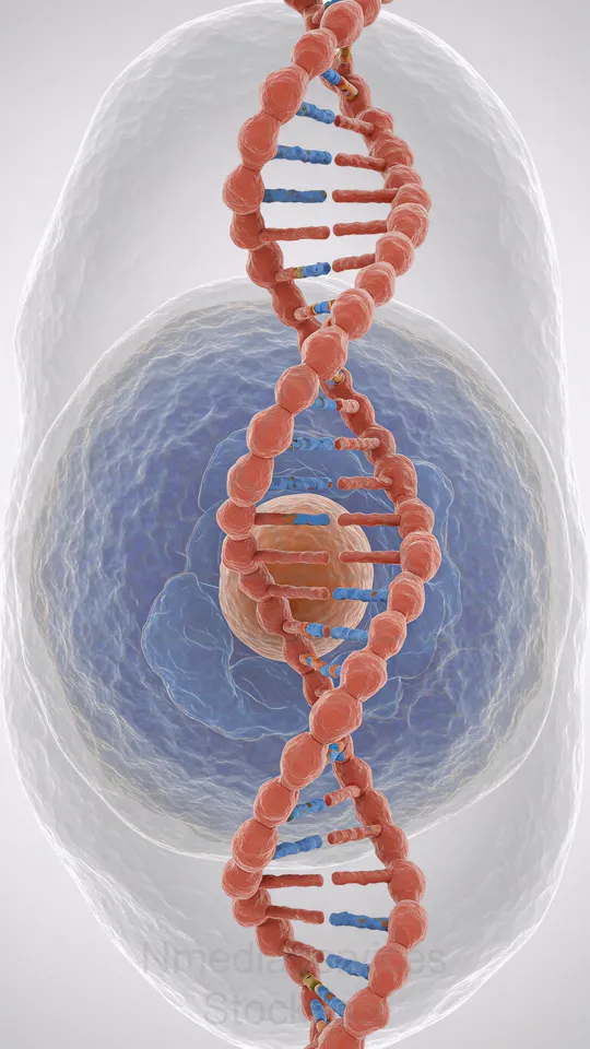
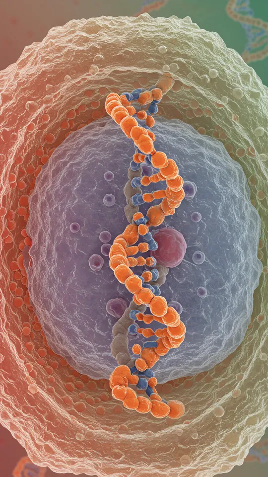
3D illustration of a cell with a prominent DNA double helix inside the nucleus.
⬇ Download / Buy on Stockflow.media
9 Micro 14 Cellular
Resolution: 11092x6050 | Format: 16:9
Fluorescent micrograph of a cellular network with red filamentous structures, green punctate signals, and blue-stained nuclei.
⬇ Download / Buy on Stockflow.media
10 Micro 14 Cellular
Resolution: 8192x8192 | Format: 1:1
Fluorescent micrograph showing red filamentous fibers with green cellular structures and blue-stained nuclei.
⬇ Download / Buy on Stockflow.media
11 Micro 14 Cellular
Resolution: 6050x11092 | Format: 9:16
Fluorescent micrograph showing red filamentous fibers with green cellular structures and blue-stained nuclei.
⬇ Download / Buy on Stockflow.media
12 Micro 14 Cellular
Resolution: 11092x6050 | Format: 16:9
Close-up of a neuron with a myelinated axon, showing signal transmission along the fiber and glowing nodes.
⬇ Download / Buy on Stockflow.media
13 Micro 14 Cellular
Resolution: 8192x8192 | Format: 1:1
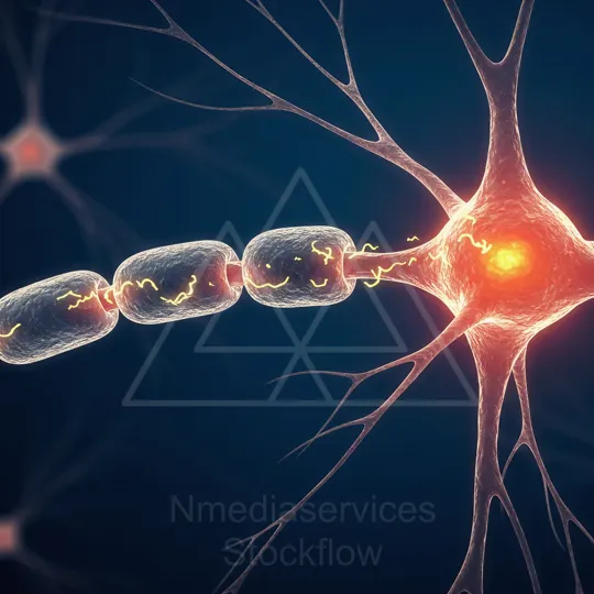
Illustration of a neuron with a myelinated axon and a glowing active region (node of Ranvier).
⬇ Download / Buy on Stockflow.media
14 Micro 14 Cellular
Resolution: 11092x6050 | Format: 16:9
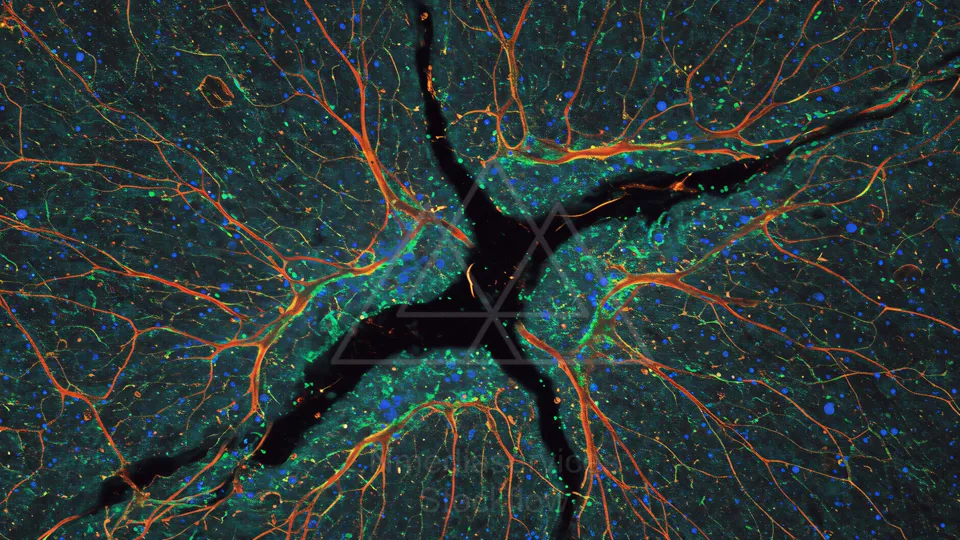
Illustration of a neuron with a myelinated axon and a glowing active region (node of Ranvier).
⬇ Download / Buy on Stockflow.media
15 Micro 14 Cellular
Resolution: 8192x8192 | Format: 1:1
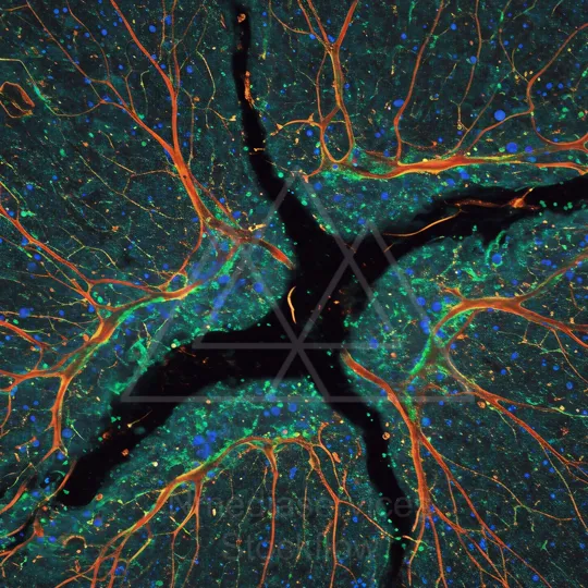
Illustration of a neuron with a myelinated axon and a glowing active region (node of Ranvier).
⬇ Download / Buy on Stockflow.media
16 Micro 14 Cellular
Resolution: 11092x6050 | Format: 16:9
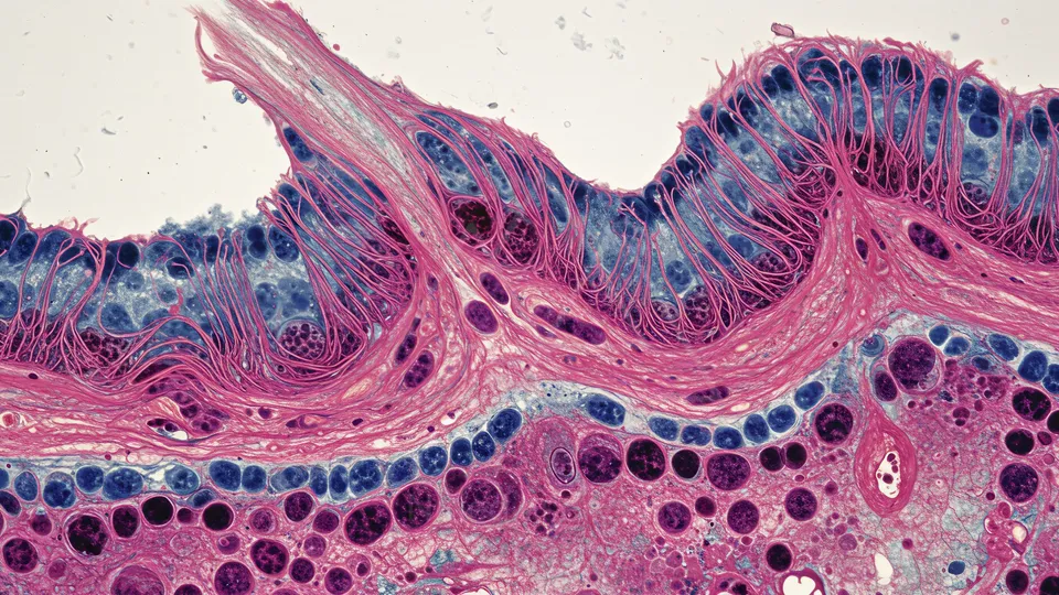
Illustration of a neuron with a myelinated axon and a glowing active region (node of Ranvier).
⬇ Download / Buy on Stockflow.media
17 Micro 14 Cellular
Resolution: 8192x8192 | Format: 1:1
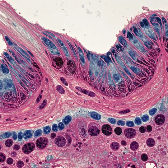
Illustration of a neuron with a myelinated axon and a glowing active region (node of Ranvier).
⬇ Download / Buy on Stockflow.media
18 Micro 14 Cellular
Resolution: 11092x6050 | Format: 16:9
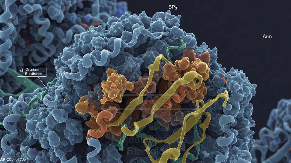
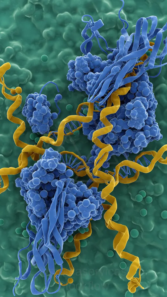
Colored electron-micrograph showing a cell surface with tangled proteins and labeled molecular components.
⬇ Download / Buy on Stockflow.media
19 Micro 14 Cellular
Resolution: 11092x6050 | Format: 16:9
Colorful DNA double helix illustration.
⬇ Download / Buy on Stockflow.media
20 Micro 14 Cellular
Resolution: 8192x8192 | Format: 1:1
Colorful 3D render of a DNA double helix with red and teal strands and colored base pairs.
⬇ Download / Buy on Stockflow.media
21 Micro 14 Cellular
Resolution: 11092x6050 | Format: 16:9
Colorful illustration of a eukaryotic cell’s interior, showing mitochondria, membranous organelles, vesicles, and surrounding particles.
⬇ Download / Buy on Stockflow.media
22 Micro 14 Cellular
Resolution: 6050x11092 | Format: 9:16
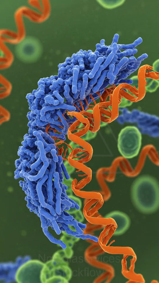
Colorized image of a blue protein complex embedded in an orange membrane helix within a cell, surrounded by green cells.
⬇ Download / Buy on Stockflow.media
23 Micro 14 Cellular
Resolution: 11092x6050 | Format: 16:9
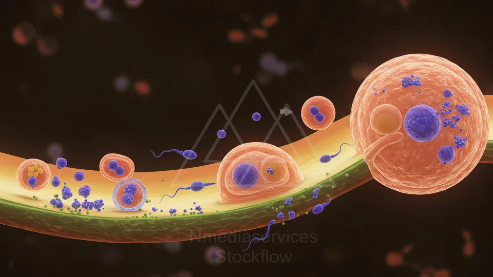
Illustration of cellular activity in a blood vessel with large orange cells, blue bacteria, and flagellated microbes.
⬇ Download / Buy on Stockflow.media
24 Micro 14 Cellular
Resolution: 11092x6050 | Format: 16:9
Fluorescent micrograph of cells with blue nuclei, green cytoskeletal network, and red actin fibers.
⬇ Download / Buy on Stockflow.media
25 Micro 14 Cellular
Resolution: 8192x8192 | Format: 1:1
Fluorescent microscopy image of a cell showing red actin filaments, green mitochondria, and blue nuclei with scattered green vesicles.
⬇ Download / Buy on Stockflow.media
26 Micro 14 Cellular
Resolution: 11092x6050 | Format: 16:9
Fluorescent microscopy image of a cell showing red actin filaments, green mitochondria, and blue nuclei with scattered green vesicles.
⬇ Download / Buy on Stockflow.media
27 Micro 14 Cellular
Resolution: 8192x8192 | Format: 1:1
Fluorescent microscopy image of a cell showing red actin filaments, green mitochondria, and blue nuclei with scattered green vesicles.
⬇ Download / Buy on Stockflow.media
28 Micro 14 Cellular
Resolution: 11092x6050 | Format: 16:9
Cross-sectional view of a blood vessel with circulating cells and cellular activity.
⬇ Download / Buy on Stockflow.media
29 Micro 14 Cellular
Resolution: 6050x11092 | Format: 9:16
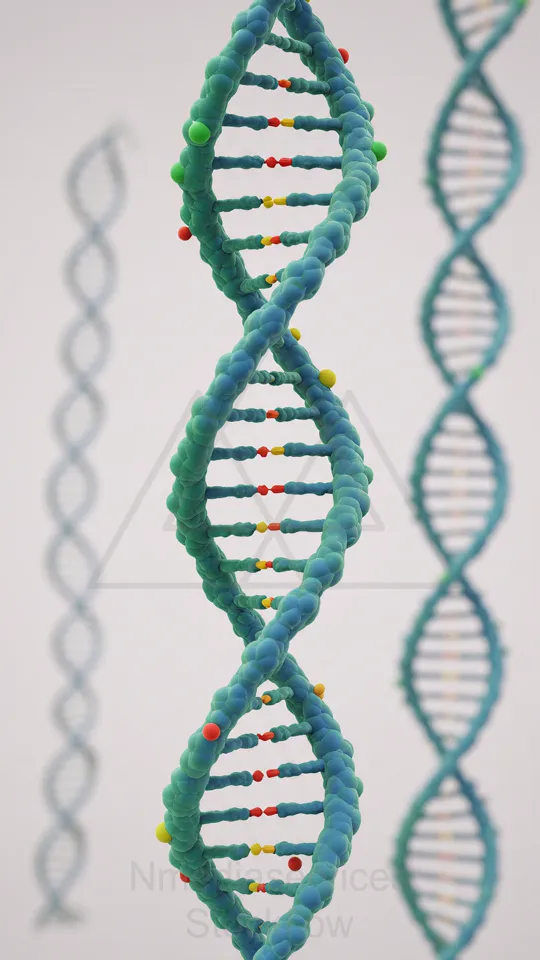
A 3D teal DNA double helix model with colorful base pairs, set against a pale background.
⬇ Download / Buy on Stockflow.media
30 Micro 14 Cellular
Resolution: 11092x6050 | Format: 16:9
intricate look at biological structures at a cellular level. Captured with advanced microscopic imaging technology
⬇ Download / Buy on Stockflow.media
31 Micro 14 Cellular
Resolution: 8192x8192 | Format: 1:1
intricate look at biological structures at a cellular level. Captured with advanced microscopic imaging technology
⬇ Download / Buy on Stockflow.media
32 Micro 14 Cellular
Resolution: 6050x11092 | Format: 9:16
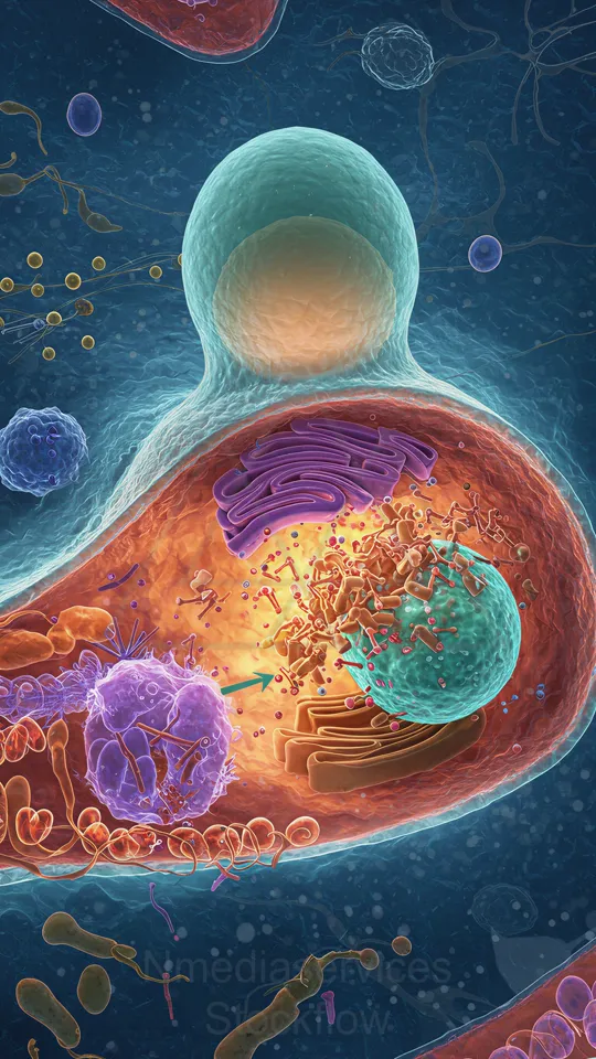
Illustration of a eukaryotic cell with visible organelles (mitochondria, endoplasmic reticulum) and surrounding extracellular environment.
⬇ Download / Buy on Stockflow.media
33 Micro 14 Cellular
Resolution: 6050x11092 | Format: 9:16
Colorized electron micrograph of a cellular membrane with large blue protein complexes and yellow cytoskeletal filaments.
⬇ Download / Buy on Stockflow.media
34 Micro 14 Cellular
Resolution: 11092x6050 | Format: 16:9
Fluorescence micrograph of epithelial tissue: blue nuclei, red cytoskeleton outlining cell borders, and green markers marking additional structures scattered throughout the tissue.
⬇ Download / Buy on Stockflow.media
35 Micro 14 Cellular
Resolution: 6050x11092 | Format: 9:16
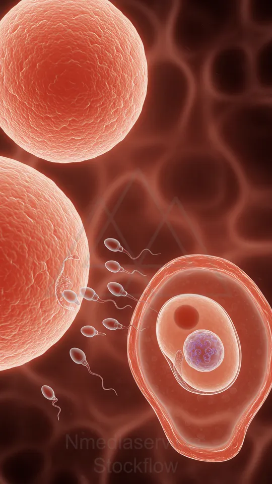
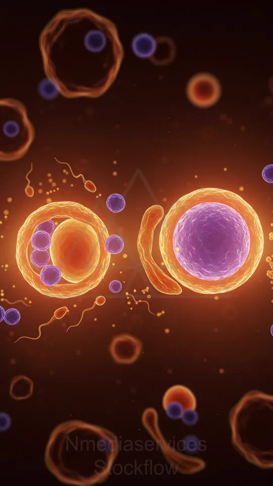
A close-up illustration of sperm approaching an ovum during fertilization, with a large egg cell and several swimming sperm surrounding it.
⬇ Download / Buy on Stockflow.media
36 Micro 14 Cellular
Resolution: 11092x6050 | Format: 16:9
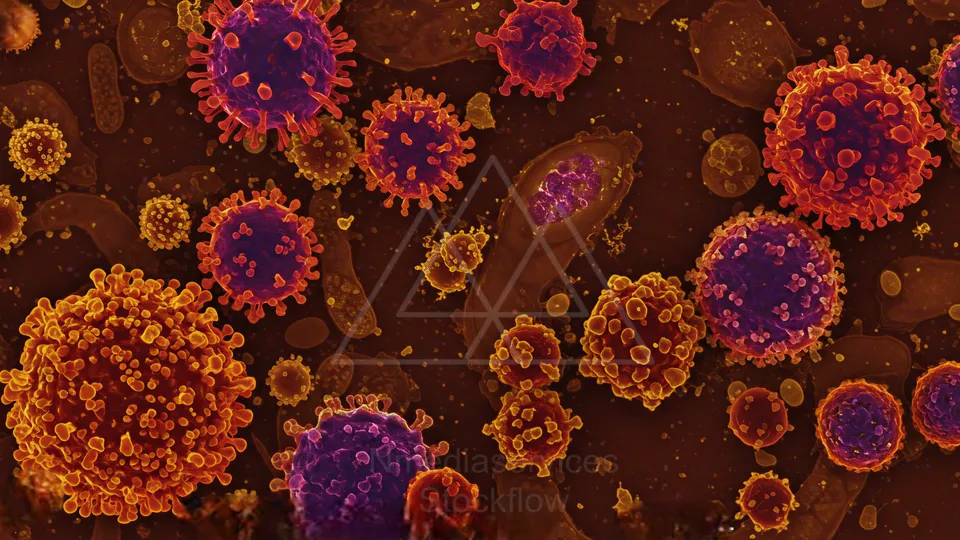
Colorized micrograph showing numerous spherical virus-like particles with spike proteins among cellular debris.
⬇ Download / Buy on Stockflow.media
37 Micro 14 Cellular
Resolution: 11092x6050 | Format: 16:9
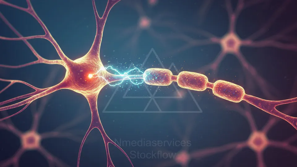
Illustration of a neuron transmitting an electrical signal across a synapse to another neuron.
⬇ Download / Buy on Stockflow.media
38 Micro 14 Cellular
Resolution: 8192x8192 | Format: 1:1
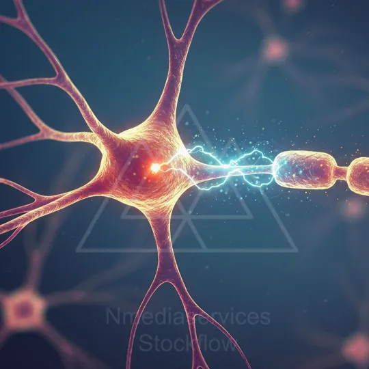
Illustration of a neuron transmitting an electrical signal along its axon.
⬇ Download / Buy on Stockflow.media
39 Micro 14 Cellular
Resolution: 11092x6050 | Format: 16:9
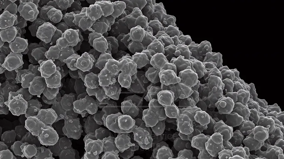
Scanning electron micrograph of densely packed, spherical bacterial cells forming a clustered colony.
⬇ Download / Buy on Stockflow.media
40 Micro 14 Cellular
Resolution: 8192x8192 | Format: 1:1
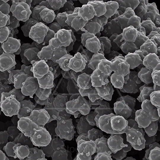
Microscopic grayscale SEM image showing a dense cluster of rounded, lobed cellular particles with textured surfaces.
⬇ Download / Buy on Stockflow.media
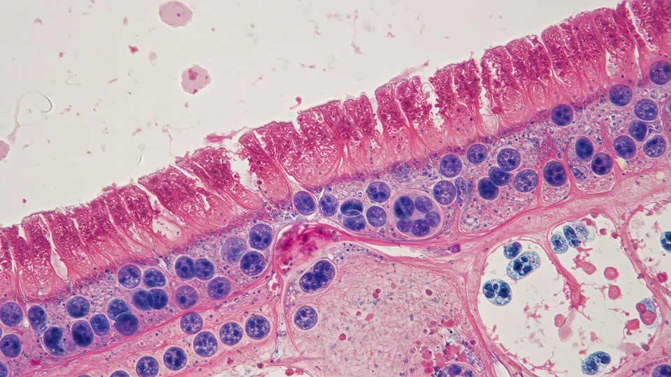
41 Micro 14 Cellular
Resolution: 6050x11092 | Format: 9:16
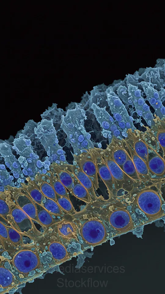
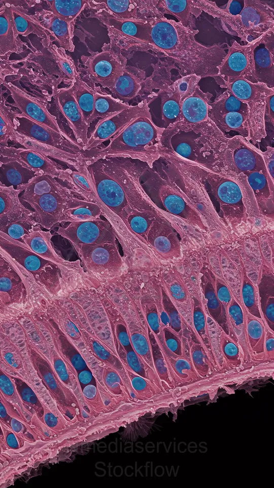
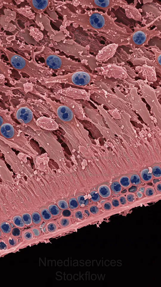
Colored scanning electron micrograph of a cellular tissue showing a row of blue-stained nuclei along a membrane with hair-like microvilli and surrounding structures.
⬇ Download / Buy on Stockflow.media
42 Micro 14 Cellular
Resolution: 6050x11092 | Format: 9:16
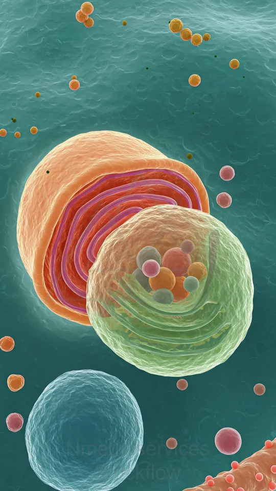
Colorful micrograph-style illustration of a cell with a prominent mitochondrion and surrounding organelles.
⬇ Download / Buy on Stockflow.media
43 Micro 14 Cellular
Resolution: 6050x11092 | Format: 9:16
High-detail illustration of a cellular membrane with a blue protein complex, yellow ribosome-like structure, and twisting red and orange protein strands.
⬇ Download / Buy on Stockflow.media
44 Micro 14 Cellular
Resolution: 6050x11092 | Format: 9:16
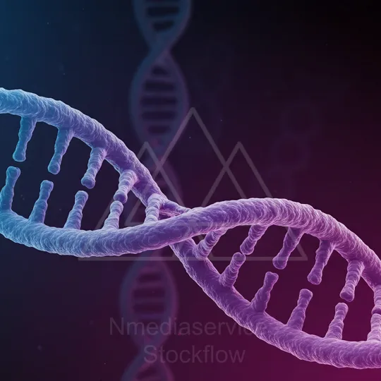
Close-up of a blue-to-purple DNA double helix strand.
⬇ Download / Buy on Stockflow.media
45 Micro 14 Cellular
Resolution: 11092x6050 | Format: 16:9
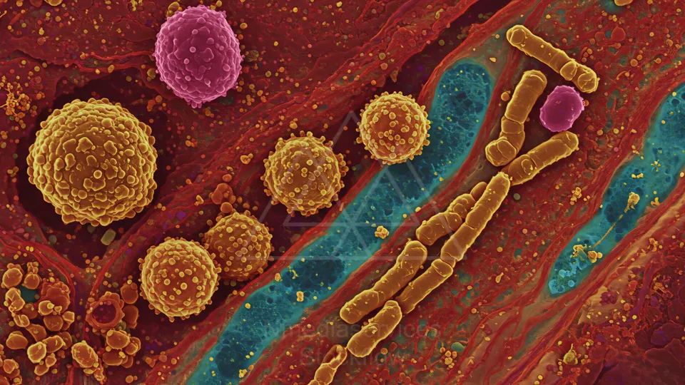
Colorized electron micrograph of round, textured cells among fibrous tissue with rod-shaped bacteria along a blue fiber.
⬇ Download / Buy on Stockflow.media
46 Micro 14 Cellular
Resolution: 8192x8192 | Format: 1:1
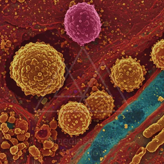
Colorized electron micrograph of round, textured cells among fibrous tissue with rod-shaped bacteria along a blue fiber.
⬇ Download / Buy on Stockflow.media
47 Micro 14 Cellular
Resolution: 6050x11092 | Format: 9:16

Close-up illustration of a green DNA double helix against a blue-purple background.
⬇ Download / Buy on Stockflow.media
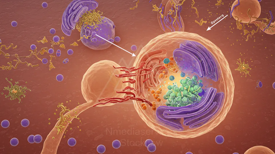
48 Micro 14 Cellular
Resolution: 11092x6050 | Format: 16:9
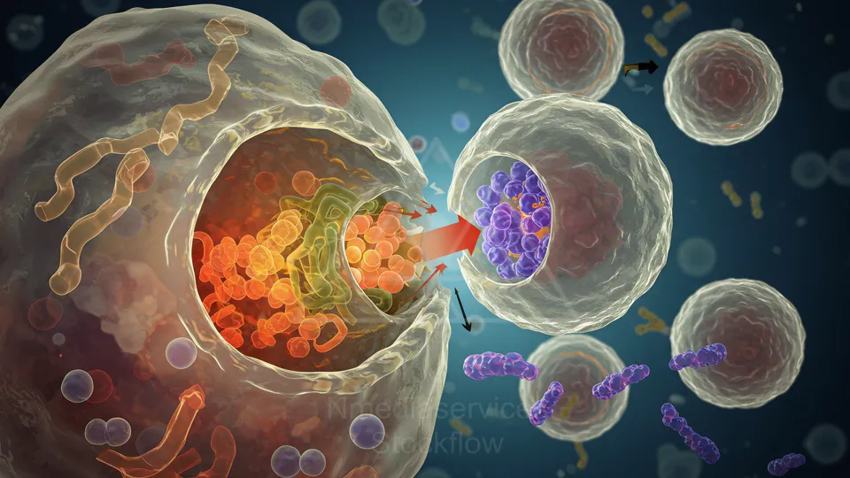
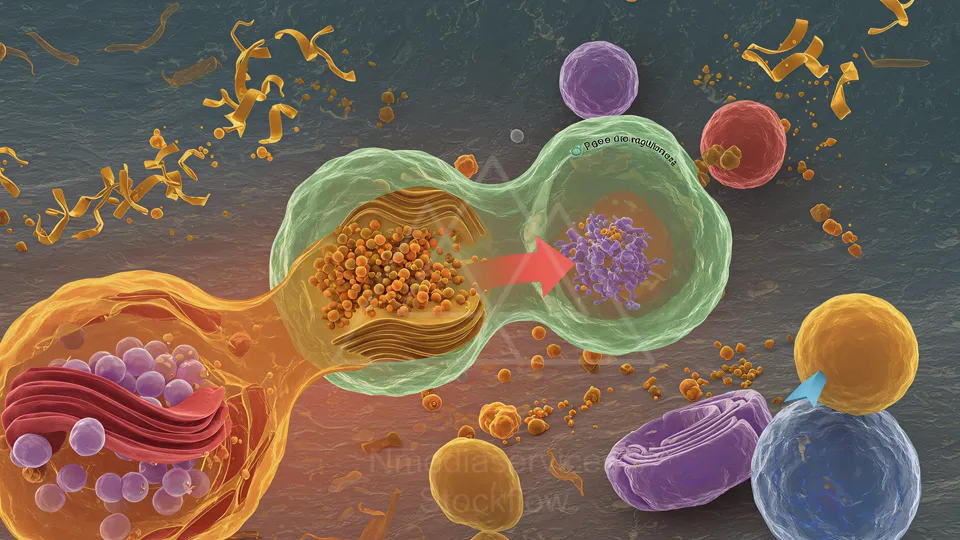
Illustration of a cell's interior with vesicles and organelles, showing a membrane-bound vesicle fusing with another cell to transfer contents.
⬇ Download / Buy on Stockflow.media
49 Micro 14 Cellular
Resolution: 8192x8192 | Format: 1:1
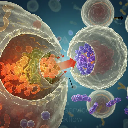
Close-up illustration of a large host cell exchanging vesicles with a neighboring cell, highlighting internal organelles and colorful vesicles in a microenvironment.
⬇ Download / Buy on Stockflow.media
50 Micro 14 Cellular
Resolution: 8192x8192 | Format: 1:1
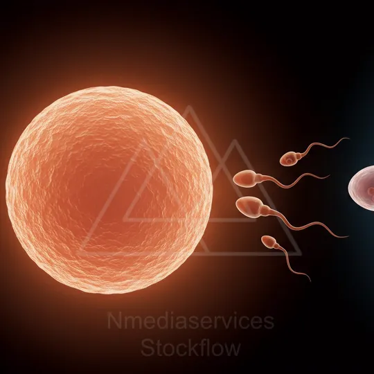
Illustration of a large orange ovum being fertilized by multiple sperm.
⬇ Download / Buy on Stockflow.media
51 Micro 14 Cellular
Resolution: 6050x11092 | Format: 9:16
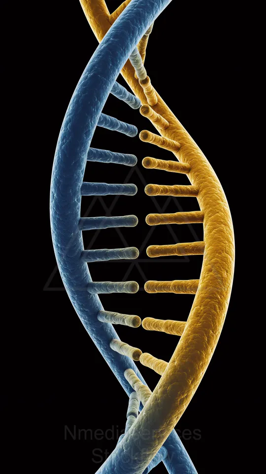
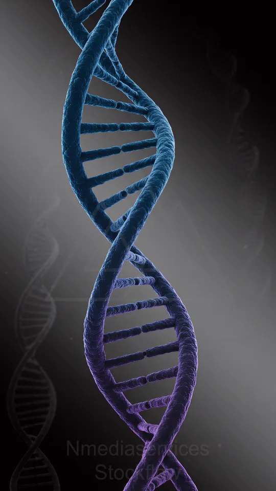
DNA double helix in blue and gold against a black background.
⬇ Download / Buy on Stockflow.media
52 Micro 14 Cellular
Resolution: 6050x11092 | Format: 9:16
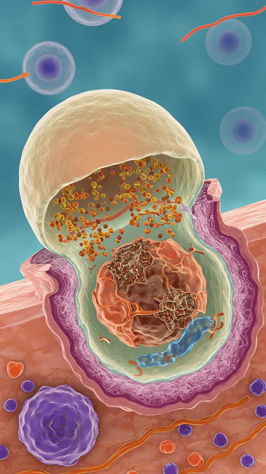
Illustration of a tissue cross-section with a large central cell showing its nucleus and organelles, surrounded by extracellular matrix and smaller cells.
⬇ Download / Buy on Stockflow.media
53 Micro 14 Cellular
Resolution: 6050x11092 | Format: 9:16
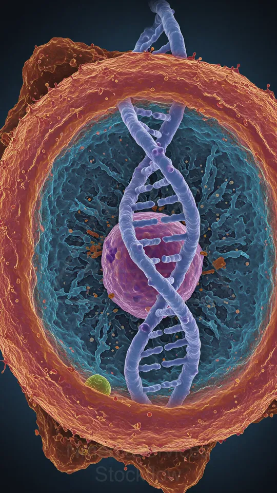
Colorized microscopic cross-section of a cell showing a DNA double helix inside the nucleus.
⬇ Download / Buy on Stockflow.media
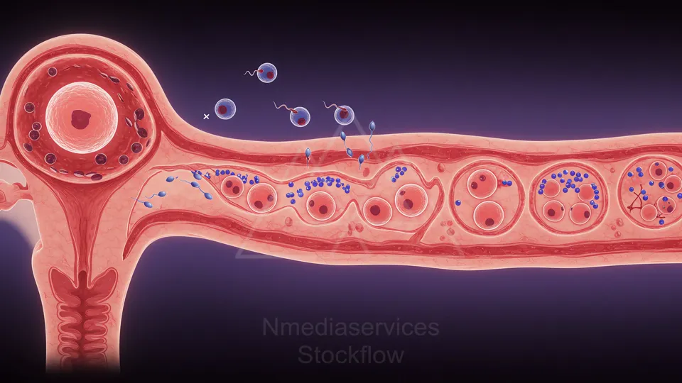
54 Micro 14 Cellular
Resolution: 6050x11092 | Format: 9:16
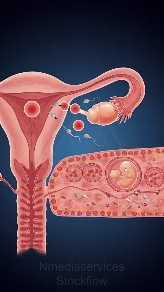
Illustration of fertilization: an egg released from the ovary into the fallopian tube, sperm swimming to meet the egg and form a zygote that implants in the uterus.
⬇ Download / Buy on Stockflow.media
55 Micro 14 Cellular
Resolution: 6050x11092 | Format: 9:16

A detailed microcosm illustration of a neuron at a synapse, showing mitochondria, vesicles, and neurotransmitter release.
⬇ Download / Buy on Stockflow.media
56 Micro 14 Cellular
Resolution: 11092x6050 | Format: 16:9

Close-up of a blue-to-pink DNA double helix.
⬇ Download / Buy on Stockflow.media
57 Micro 14 Cellular
Resolution: 8192x8192 | Format: 1:1
Microscopic view of a purple-blue DNA double helix against a dark gradient background.
⬇ Download / Buy on Stockflow.media
58 Micro 14 Cellular
Resolution: 11092x6050 | Format: 16:9
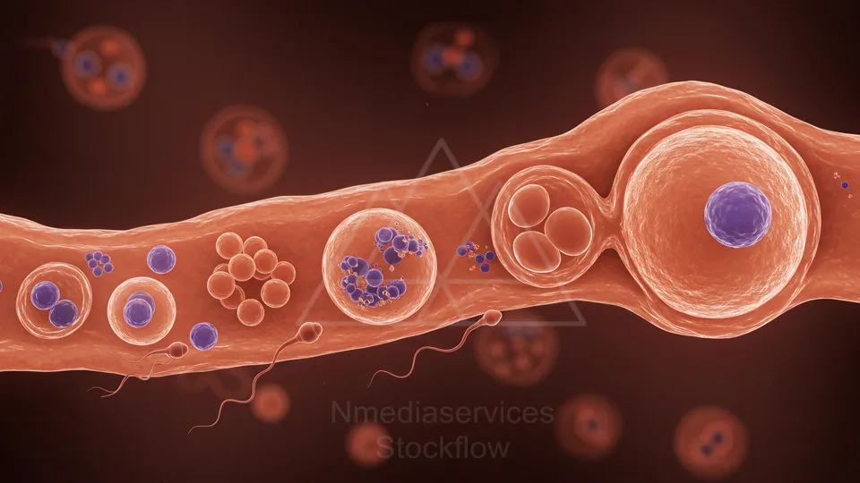
Colorful illustration of cellular elements during fertilization, showing sperm approaching an egg and early embryo development.
⬇ Download / Buy on Stockflow.media
59 Micro 14 Cellular
Resolution: 8192x8192 | Format: 1:1
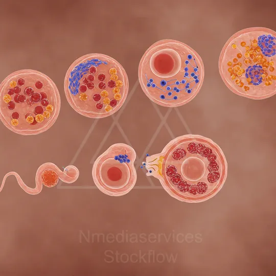
Illustration of several magnified circular cellular structures showing different organelles and components.
⬇ Download / Buy on Stockflow.media
60 Micro 14 Cellular
Resolution: 8192x8192 | Format: 1:1
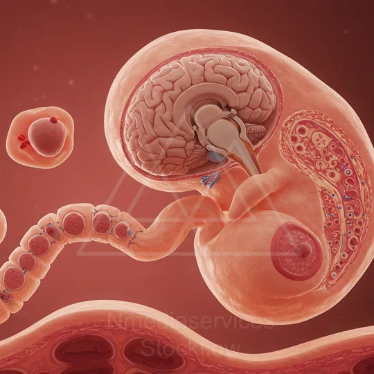
In utero fetus illustration with developing brain and spine, connected by the umbilical cord to the placenta.
⬇ Download / Buy on Stockflow.media
61 Micro 14 Cellular
Resolution: 6050x11092 | Format: 9:16

3D rendering of a cell interior showing blue tubular filaments forming a tangled network inside a pinkish cell membrane.
⬇ Download / Buy on Stockflow.media
62 Micro 14 Cellular
Resolution: 11092x6050 | Format: 16:9
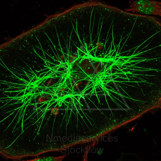
Fluorescence micrograph of a cell showing a dense green tubular cytoskeleton spanning the cytoplasm, with a red actin cortex outlining the cell and green intracellular granules.
⬇ Download / Buy on Stockflow.media
63 Micro 14 Cellular
Resolution: 8192x8192 | Format: 1:1
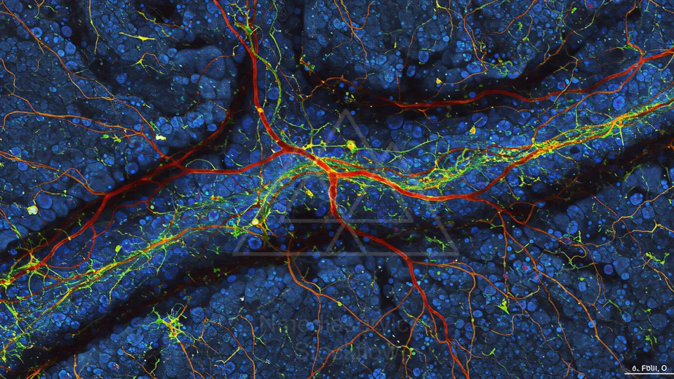
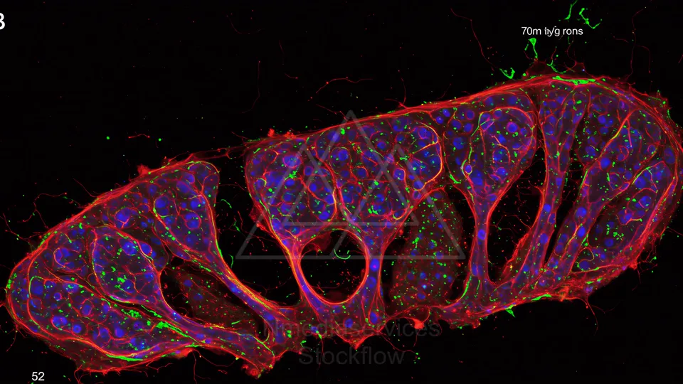
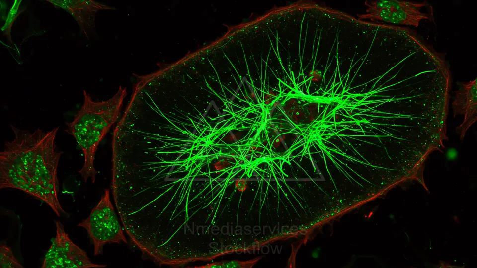
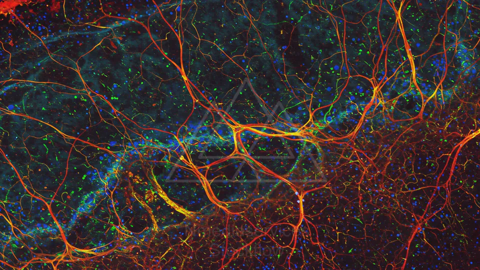
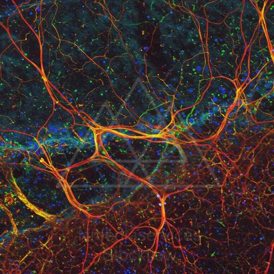
Fluorescent micrograph of neurons with extensive green axonal processes in a tissue slice.
⬇ Download / Buy on Stockflow.media
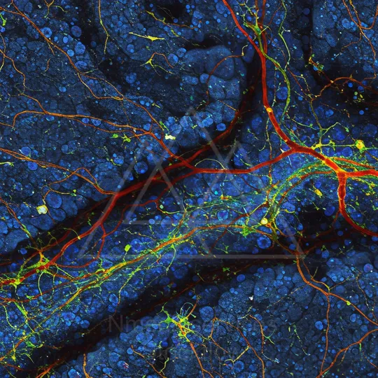
64 Micro 14 Cellular
Resolution: 6050x11092 | Format: 9:16
Fluorescent micrograph of neurons with extensive green axonal processes in a tissue slice.
⬇ Download / Buy on Stockflow.media
65 Micro 14 Cellular
Resolution: 11092x6050 | Format: 16:9
A detailed illustration of cellular activity inside a vessel, showing vesicles, clusters of cells, and motile, sperm-like structures.
⬇ Download / Buy on Stockflow.media
66 Micro 14 Cellular
Resolution: 11092x6050 | Format: 16:9
Close-up illustration of a neuron with interconnected axons and blue-green myelin sheaths glowing against a dark background.
⬇ Download / Buy on Stockflow.media
67 Micro 14 Cellular
Resolution: 8192x8192 | Format: 1:1
Glowing neuron with elongated axon and branching dendrites.
⬇ Download / Buy on Stockflow.media
68 Micro 14 Cellular
Resolution: 11092x6050 | Format: 16:9
Colorful 3D rendering of clustered cells in blue, green and orange tones.
⬇ Download / Buy on Stockflow.media
69 Micro 14 Cellular
Resolution: 8192x8192 | Format: 1:1
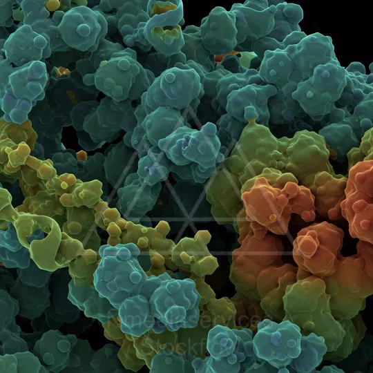
Vivid 3D rendering of colorful cellular clusters in teal, blue, green, yellow, and orange.
⬇ Download / Buy on Stockflow.media
70 Micro 14 Cellular
Resolution: 11092x6050 | Format: 16:9
Colorful 3D illustration of a cell with vesicles and organelles undergoing membrane fusion and intracellular transport.
⬇ Download / Buy on Stockflow.media
71 Micro 14 Cellular
Resolution: 11092x6050 | Format: 16:9
Colorful 3D illustration of a cell with vesicles and organelles undergoing membrane fusion and intracellular transport.
⬇ Download / Buy on Stockflow.media
72 Micro 14 Cellular
Resolution: 8192x8192 | Format: 1:1
Colorful 3D illustration of a cell with vesicles and organelles undergoing membrane fusion and intracellular transport.
⬇ Download / Buy on Stockflow.media
73 Micro 14 Cellular
Resolution: 6050x11092 | Format: 9:16
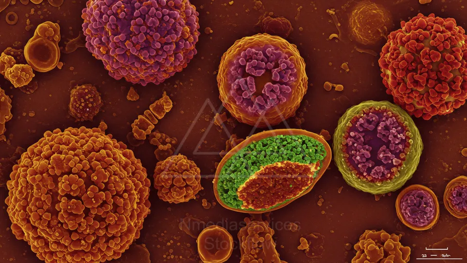
Color-enhanced micrograph of cells with orange rings and purple interiors, surrounded by smaller purple spheres and stringy structures against a warm amber background.
⬇ Download / Buy on Stockflow.media
74 Micro 14 Cellular
Resolution: 11092x6050 | Format: 16:9
Close-up of a cell membrane showing blue membrane proteins, a yellow receptor cluster, and orange organelles within a textured, greenish surface.
⬇ Download / Buy on Stockflow.media
75 Micro 14 Cellular
Resolution: 8192x8192 | Format: 1:1
Colorful 3D render of a cell membrane scene with a yellow receptor and blue protein channels embedded in a lipid bilayer, surrounded by various molecules.
⬇ Download / Buy on Stockflow.media
76 Micro 14 Cellular
Resolution: 11092x6050 | Format: 16:9

Close-up illustration of a blue-purple DNA double helix with orange base pairs.
⬇ Download / Buy on Stockflow.media
77 Micro 14 Cellular
Resolution: 8192x8192 | Format: 1:1

Close-up of a blue-purple DNA double helix with orange base pairs.
⬇ Download / Buy on Stockflow.media
78 Micro 14 Cellular
Resolution: 11092x6050 | Format: 16:9
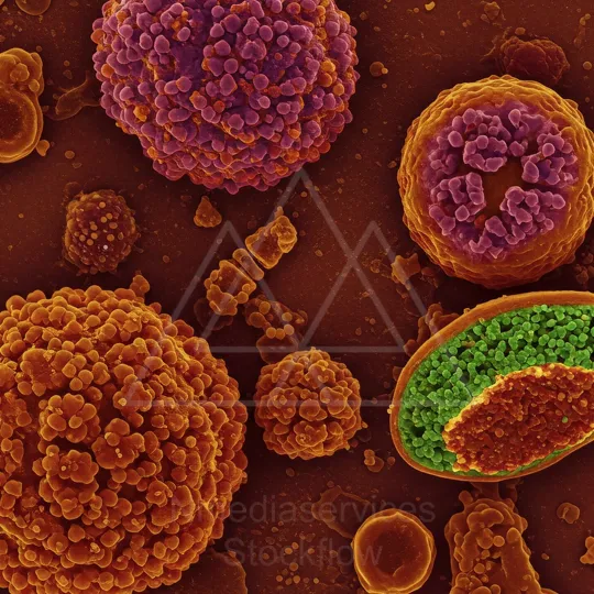
Colorful microscopic view of diverse spherical cells with spiky surfaces
⬇ Download / Buy on Stockflow.media
79 Micro 14 Cellular
Resolution: 8192x8192 | Format: 1:1
Colorful microscopic view of diverse spherical cells with spiky surfaces
⬇ Download / Buy on Stockflow.media
80 Micro 14 Cellular
Resolution: 11092x6050 | Format: 16:9
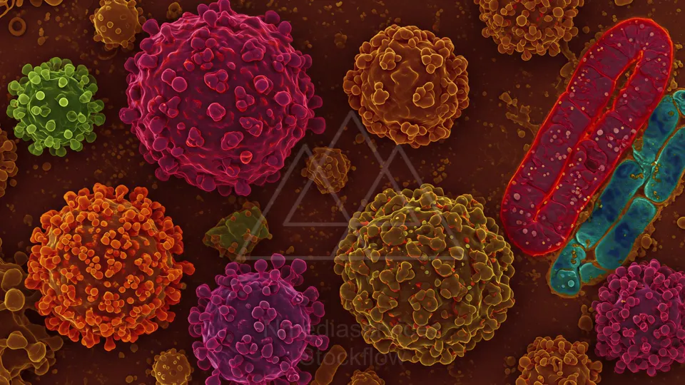
Colorful microscopic view of diverse spherical cells with spiky surfaces and a red elongated organelle among them.
⬇ Download / Buy on Stockflow.media
81 Micro 14 Cellular
Resolution: 8192x8192 | Format: 1:1

Colorful 3D render of multiple spherical cellular particles with surface projections against a dark background.
⬇ Download / Buy on Stockflow.media
1 Micro 00
Resolution: 6050x11092 | Format: 9:16
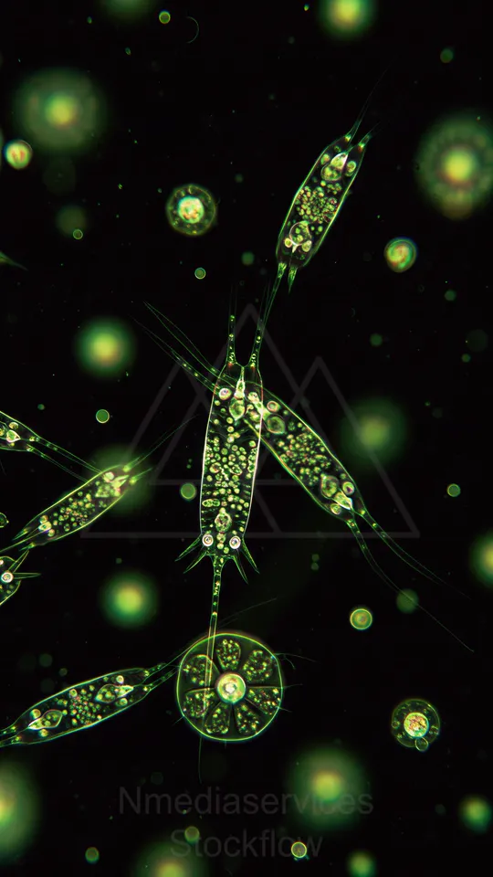
Fluorescent green micrograph of elongated, segmented aquatic microorganisms with internal granules and fine thread-like appendages.
⬇ Download / Buy on Stockflow.media
01 Cellular
Resolution: 2160x2160 | Format: 1:1
A high-detail micrograph-style scene of a cell membrane with twisting protein structures, colorful receptors and molecular machinery, illustrating cellular interactions and signaling on a miniature, vivid biological landscape.
⬇ Download / Buy on Stockflow.media
02 Cellular
Resolution: 2160x3840 | Format: 9:16
A detailed, micro-scale illustration of nerve cells and myelinated axons forming a neural network, highlighting connections, synapses, and cellular structure in cool blue tones across the image.
⬇ Download / Buy on Stockflow.media
03 Cellular
Resolution: 2160x3840 | Format: 9:16
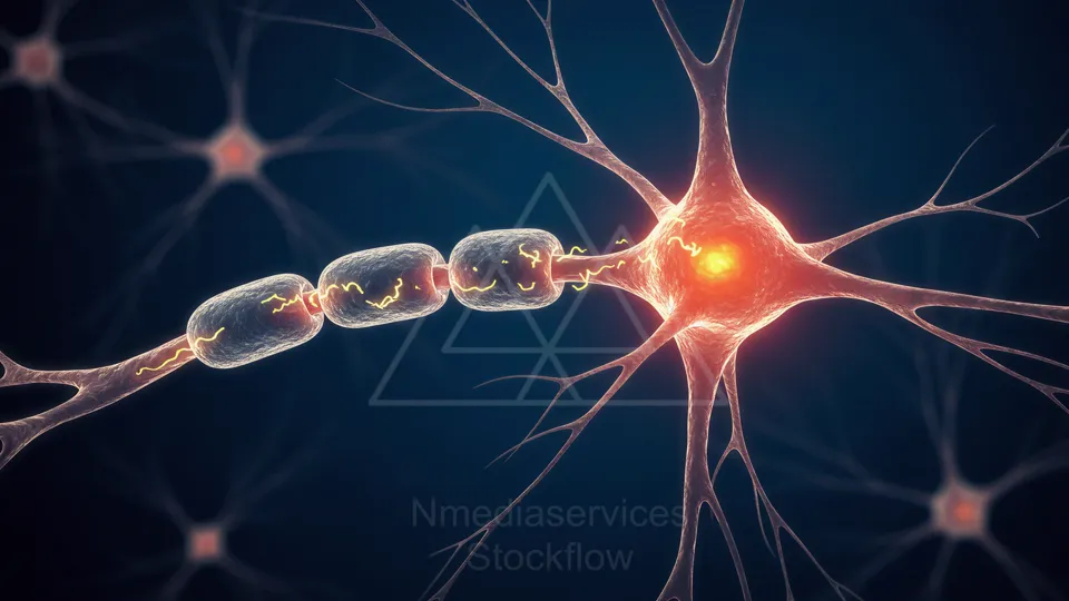
A vivid illustration of a neuron with a glowing cell body and a chain of myelinated axon segments, highlighting electrical signals traveling along the neural pathway.
⬇ Download / Buy on Stockflow.media
04 Cellular
Resolution: 2160x3840 | Format: 9:16
A vivid micrographic view inside a blood vessel, showing numerous red blood cells flowing past white blood cells and tiny particles, illustrating the dynamic environment of the circulatory system.
⬇ Download / Buy on Stockflow.media
05 Cellular
Resolution: 2160x3840 | Format: 9:16
A glowing neuron stretches its axon, with bright cell bodies along the chain and shimmering electrical signals coursing through yellow myelin. The scene highlights neural connectivity and communication.
⬇ Download / Buy on Stockflow.media
06 Cellular
Resolution: 2160x3840 | Format: 9:16
A vivid view inside a blood vessel shows flowing red blood cells mingling with blue-white immune cells and scattered platelets, illustrating circulation, cellular interactions, and the dynamic inner environment of the bloodstream.
⬇ Download / Buy on Stockflow.media
07 Cellular
Resolution: 2160x3840 | Format: 9:16
Vibrant microscopic view of a neuron with glowing myelinated axons transmitting electric signals, synapses sparking at the nodes, highlighting neural communication and the intricate network of brain cells.
⬇ Download / Buy on Stockflow.media
20 Cellular
Resolution: 3840x2176 | Format: 16:9
A highly detailed microscopic view of a cell membrane showing colorful proteins and molecular machinery at work, illustrating how receptors, channels, and signaling elements organize on the cellular surface.
⬇ Download / Buy on Stockflow.media
21 Cellular
Resolution: 1064x1064 | Format: 1:1
Fluorescent microscopy of cellular structures revealing blue nuclei, green cytoplasm outlines, and bright red actin filaments radiating from focal centers, illustrating cytoskeleton organization in cells under high resolution.
⬇ Download / Buy on Stockflow.media
22 Cellular
Resolution: 40x2160 | Format: 16:9
Fluorescent microscopy image showing two adjacent cells with a vivid actin network (red) extending across the boundary, green cytoplasm, and blue-stained nuclei illustrating cytoskeletal organization and intercellular contact.
⬇ Download / Buy on Stockflow.media
23 Cellular
Resolution: 1088x1088 | Format: 1:1
A high-resolution 3D rendering of a cell membrane showing blue transmembrane proteins and yellow signaling molecule clusters amid a green lipid bilayer, illustrating molecular interactions at the cell surface.
⬇ Download / Buy on Stockflow.media
24 Cellular
Resolution: 2160x3840 | Format: 9:16
A vivid microscopic view inside a blood vessel, where numerous red blood cells flow past blue-white immune cells and various platelets, highlighting the dynamic, bustling environment of circulating blood.
⬇ Download / Buy on Stockflow.media
25 Cellular
Resolution: 2160x3840 | Format: 9:16
A vivid microscopic view of a blood vessel showing circulating red blood cells and white blood cells amid plasma, illustrating cellular components of the circulatory system.
⬇ Download / Buy on Stockflow.media
26 Cellular
Resolution: 3840x2160 | Format: 16:9
A vivid microscopic view of a cell membrane with diverse colorful proteins and molecular machinery, illustrating how receptors and channels organize on the surface in intricate, dynamic detail.
⬇ Download / Buy on Stockflow.media
06 Microscopic
Resolution: 3840x2160 | Format: 16:9
Close-up view of a virus particle looming over a cell membrane, with colorful molecules and membranous structures, illustrating viral entry and cellular interactions at a tiny scale.
⬇ Download / Buy on Stockflow.media
07 Microscopic
Resolution: 2160x2160 | Format: 1:1
A vivid, macro view of cellular activity and microstructures in glowing magenta-orange tones, hinting at cells communicating and entering new phases within a dynamic biological environment.
⬇ Download / Buy on Stockflow.media
08 Microscopic
Resolution: 2160x2160 | Format: 1:1
Entering a micro cell universe: vivid swirling cytoplasm around a glowing nucleus reveals a dynamic, immersive internal world.
⬇ Download / Buy on Stockflow.media
09 Microscopic
Resolution: 2160x2160 | Format: 1:1
Glowing green microscopic organisms float in a dark, watery world, their translucent bodies revealing internal structures as delicate filaments drift and intertwine in a shimmering underwater scene.
⬇ Download / Buy on Stockflow.media
10 Microscopic
Resolution: 3840x2160 | Format: 16:9
A vibrant microscopic scene shows glowing green filamentous organisms drifting in dark water, their bright cells linked by fine blue fibers, creating a surreal underwater network of life.
⬇ Download / Buy on Stockflow.media